
Introduction
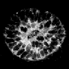
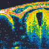
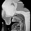
Optical projection tomography (OPT) microscopy is a new technique which allows the 3D imaging of biological specimen over 1 cm across. It was initially developed with the hope of enabling accurate measurement of 3D shapes. However, it has proven to be a fast method for scanning the 3D distributions of gene expression patterns during embryo development. In particular it has two important advantages over confocal microscopy: it can image much larger specimen, and it can image non-fluorescent specimen. This means that BCIP/NBT in-situs or LacZ staining can be mapped in 3D. Fluorescent signals can also be imaged, allowing for the mapping of multiple protein distributions within the same tissue. It also has a number of advantages over microscopic magnetic resonance imaging (mMRI), including speed, resolution, the ability to image commonly-used stains, and cost.OPT is based on the principle of projection tomography, just like X-ray CT scanners. This is in contrast to techniques such as confocal microscopy or optical coherence tomography (OCT), both of which are examples of section tomography.
Links:
- Background to the invention - The need for 3D data
- What's in a name? - A definition of OPT and tomography
- How OPT compares to other techniques - an overview
|
|
|

|