
The eHistology Resource with Kaufman Annotations and Coronals Supplement
|
 |
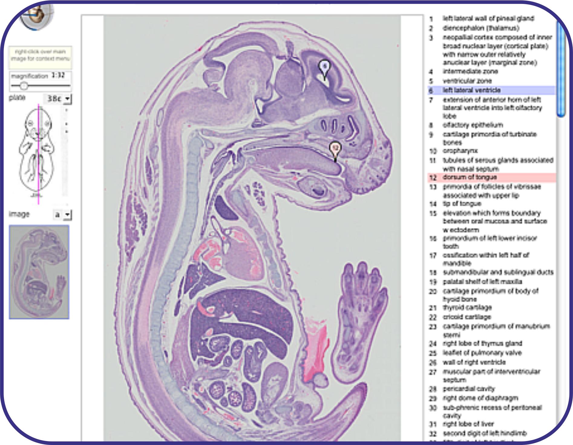
The original paper publication of "The Atlas of Mouse Development" by M.H.Kaufman (1992, Academic Press, London) is the definitive work for mouse developmental anatomy. The original figures were generated from black and white photographs of the H&E stained sections painstakingly annotated by hand by Professor Kaufman. Here we have re-digitised, in high-resolution and colour, the original histology sections on glass slides and now make the new set of images freely available with a zoom-viewer. In collaboration with Elsevier these new images show the annotation from the original paper atlas and are linked through to other mouse resources such as the eMouseAtlas and MGI/GXD. The full book content is likely to be made available via online material and an eBook from Elsevier using these images and annotations. Note: all images and associated metadata are available for free download and can be used for any purpose provided appropriate citation is made. |
 |
 |
A supplement to the original Atlas of Mouse Development has been published and the coronal histology images of the original have been extended to more stages and presented online as full sections through the embryo. The annotations from the book are presented in the same way as labels with markers in the zoomable image. 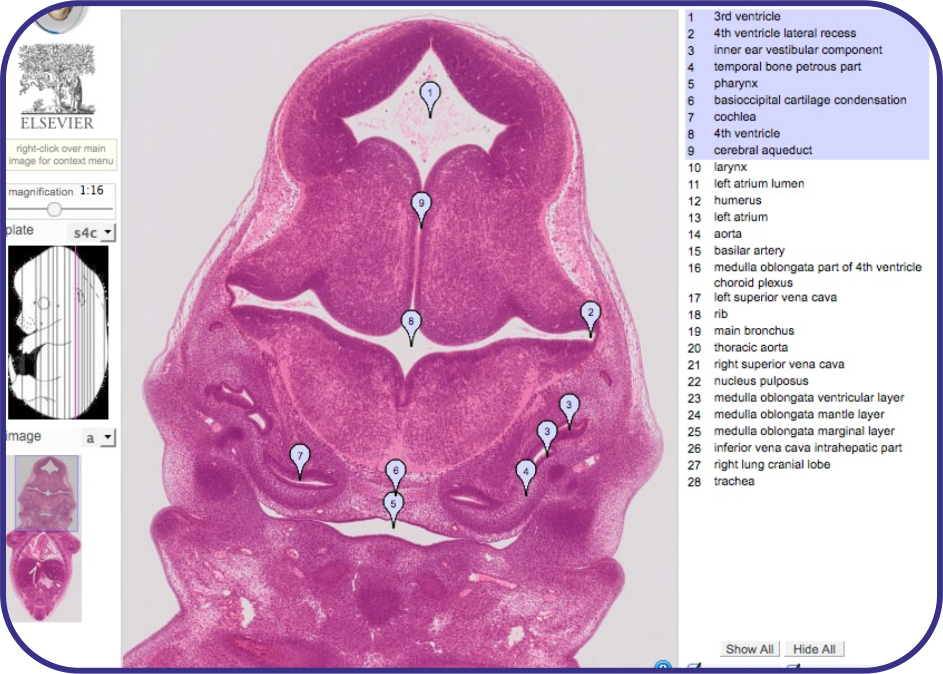 |
|
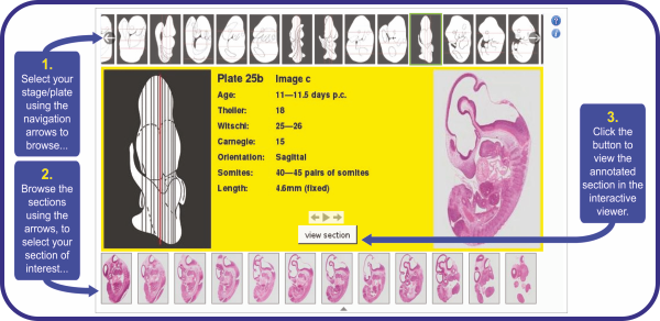
The online view of each image can be found using the eHistology interactive image index. Selecting the plate from the film-strip at the top presents a view of all the section images available for that embryo. Details of the stage and and approximate location of the section within the embryo are provided in the central panel using a composition from the original diagrams in the book. In due course this index page will include a search option, allowing users to search for particular terms within the resource. This will also allow the section date here to be linked through from other parts of the eMouseAtlas resource including the 3D models, anatomy ontology and gene-expression database. |
 |
|
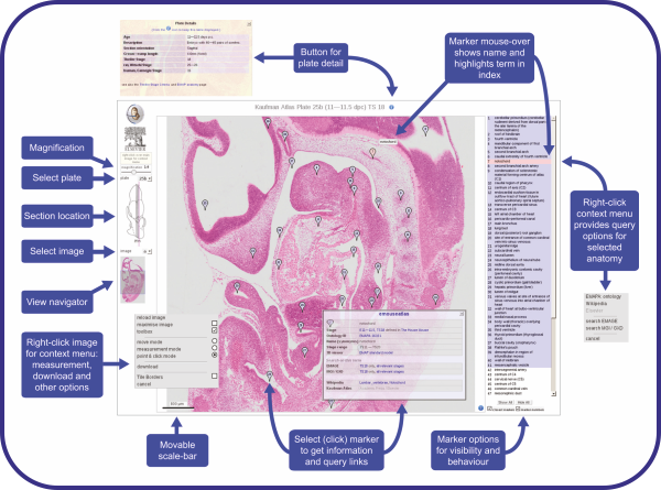
The image viewer provides a "pan and zoom" interface to deliver any resolution views of the histology through to the cellular resolution captured using a x20 objective. Controls and tools are provided for:
|
 |